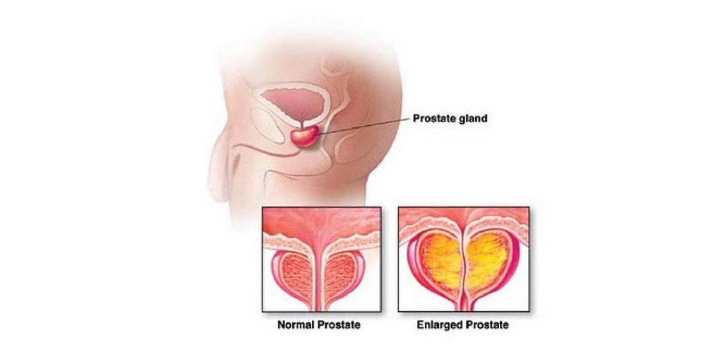
Nursing Path

CARING is the essence of NURSING. -Jean Watson

Nursing Path

Knowing is not enough, we must APPLY. Willing is not enough, we must DO. -Bruce Lee

Nursing Path

Treat the patient as a whole, not just the hole in the patient.

Nursing Path

Success is not final. Failure is not fatal. It is the courage to continue that counts. -Winston Churchill

Nursing Path

A problem is a chance for you to do your best. -Duke Ellington

pneumonia

Bladder Cancer Management and Nursing Care Plan

Description 
    Bladder Cancer
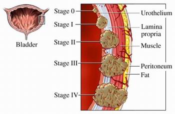
  1. Bladder cancer is papillomatous growth in the bladder urothelium that undergo malignant changes and that may infiltrate the bladder wall.
  2. Predisposing factors include cigarette smoking, exposure to industrial chemicals and exposure to radiation.
  3. Common signs of metastasis include the liver, bones and lungs
  4. As the tumor progresses can extend to the rectum, vagina and retro- peritoneal structures.
Assessment 
  • Painless hematuria
  • Dysuria
  • Gross hematuria
  • Obstruction of urine flow
  • Development of fistula ( urine from the vagina, fecal material in the urine)
Diagnostic Evaluation
Biopsies of the tumor and adjacent mucosa are definitive, but the following procedures are also used:
  • Cystoscopy, biopsy of tumor and adjacent mucosa
  • Excretory urography
  • Computed CT Scan
  • Ultrasonography
  • Bimanual examination by anesthesia
  • Cytologic evaluation of fresh urine and saline bladder washings
Molecular assays, bladder tumor antigens, adhesion molecules and others are being studied.
Primary Nursing Diagnosis
  • Risk for altered urinary elimination related to the obstruction of urinary flow
Medical Management
Radiation
  1. Most bladder cancer are poorly radio sensitive and require high doses of radiation
  2. Radiation therapy is more acceptable for advance disease that cannot be eradicated by surgery.
  3. Palliative radiation maybe used to relieve pain and bowel obstruction and control potential hemorrhage and leg edema cause by venous or lymphatic obstruction.
  4. Intracavitary radiation maybe prescribed which protect adjacent tissues.
  5. External radiation combined with chemotherapy or surgery maybe prescribed because the external radiation alone maybe ineffective.
  6. Complications of radiations:
    1. A bacterial cystitis
    2. Proctitis
    3. Fistula formation
    4. Ileitis or colitis
    5. Bladder ulceration and hemorrhage
Chemotherapy
1. Intravesical instillation
  • An alkylating chemotherapeutic agent is instilled into the bladder
  • This method provides an concentrated topical treatment with little systemic absorption
  • Chemotherapeutic agents used may include thiotepa, mitomycin (Mutamycin), doxorubicin (Adriamycin), cyclophosphamide (Cytoxan), and bacille Calmette-Guerin.
  • The medication is injected into a urethral catheter and retain for two hours.
  • Following instillation, the clients position is rotated every 15 to 30 minutes, starting in the supine position to avoid lying on full bladder.
  • After 2 hours, the client voids in a sitting position and is instructed to increase fluids to flash the bladder.
  • Treat the urine as biohazard and send to radioisotope laboratory for monitoring.
  • For 6 hours following intravesical therapy, disinfect the toilet with household bleach after the client has voided.
2. Systemic chemotherapy
  • Systemic chemotherapy is used to treat inoperable or late tumors.
  • Agents used may include, cysplatin (Platinol), doxorubicin (Adremycin), cyclophospamide (Cytosan), methotrexate (Folex) and Pyridoxine
3. Complications of chemotherapy
  • Bladder irritation
  • Hemorrhagic cystitis
Surgical Interventions
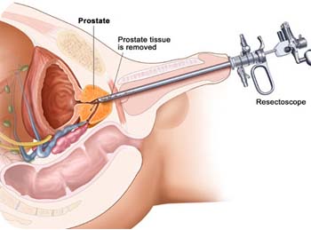
1. Transurethral resection of the bladder
  • Local resection and fulguration ( destruction of tissue by electrical current through electrodes place in direct contact with the tissue)
  • Perform for early tumor for cure or for inoperable tumors for palliation.
2. Partial Cystectomy
  • Partial cystectomy is the removal of up to half of the bladder
  • The procedure is done for early tumors and for clients who cannot tolerate radical cystectomy.
  • During the initial postoperative period bladder capacity is reduced greatly to about 60 mL; however, as the bladder tissue expand, the capacity increases to 200 -400 mL.
  • Maintenance of a continuous output of urine following surgery is critical to prevent bladder distention and stress on the suture line.
  • A urethral catheter and a suprapubic catheter maybe in place, in the suprapubic catheter maybe left in place for 2 weeks until healing occurs.
3. Cystectomy and urinary diversion
  • The procedure involves removal of the bladder and urethra in the women, and the bladder, the urethra, and usually the prostrate and seminal vesicles in men.
  • When the bladder and urethra are remove, permanent urinary diversion is required.
  • The surgery meybe performed into stages if the tumor is expensive, with the creation of the urinary diversion first and the cystectomy several weeks later.
  • If a radical cystectomy is performed lower extremity lymphedema may occur as a result of lymp node dissection, and impotence may occur in the may client.
4. Ileal conduit
  • The ileal conduit also is called ureteroileostomy or Bricker’s procedure.
  • Ureters are implanted into a segment of the ileum, with the formation of an abdominal stoma.
  • The urine flows into the conduit and is propelled continually out through the stoma by peristalsis.
  • The client is required to wear an appliance over the stoma to collect the urine.
  • Complications include obstruction, pyelonephritis, leakage at the anastomosis site, stenosis, hydronephrosis, calculuses, skin irritation and ulceration, and stomal defects.
5. Kock pouch
  • The Koch pouch is a continent internal ileal reservoir created from a segment of the ileum and ascendingcolon.
  • The ureters are implanted into the side of the reservoir, and a special nipple valve is constructed to attach the reservoir to the skin.
  • Postoperatively, the client will have a 24 to 26 Foley catheter in place to drain urine continuously until the pouch has healed.
  • The catheter is irrigated gently with NS to prevent obstruction from mucus or clots.
  • Following removal of the catheter, the client is instructed in how to self-catheterize and to drain the reservoir at 4 to 6 hour intervals.
6. Indiana pouch
  • A continent reservoir is created from the ascending colon and terminal ileum, making a pouch larger than the Koch pouch.
  • Postoperatively, the client will have a 24 to 26 Foley catheter inplace to drain urine continuously until the pouch has healed.
  • The Foley catheter is irrigated gently with NS to prevent obstruction from mucus or clots.
  • Following removal of the Foley catheter, the client is instructed in how to self-catheterize and to drain the reservoir at 4 to 6 hour intervals.
7. Creation of a neobladder
  • Creation of a neobladder is similar to the creation of an internal reservoir, with the difference being that instead of emptying through an abdominal stoma, the bladder empties through a pelvic outlet into the urethra.
  • The client empties the neobladder by relaxing the external sphincter and creating abdominal pressure or by intermittent self- catheterization.
8. Percutaneous nephrostomy or pyelostomy
  • These procedures are used when the cancer is inoperable to prevent obstruction.
  • The procedures involve a percutaneous or surgical insertion of a nephrostomy tube into the kidney for drainage.
  • Nursing interventions involves stabilizing the tube to prevent dislodgement and monitoring output.
9. Ureterostomy
  • Ureterostomy may be performed as a palliative procedure if the ureters are obstructed by the tumor.
  • The ureters are attached to the surface of the abdomen, where the urine flows directly into a drainage appliance without a conduit.
  • Potential problems include infection, skin irritation,and obstruction to urinary flow as a result of strictures at the opening.
10. Vesicostomy
  • The bladder is sutured to the abdomen, and a stoma is created in the bladder wall.
  • The bladder empties through the stoma.
Pharmacologic Intervention
  • Chemotheraphy with a combination of methotrexate, 5-fluorouracil (5-FU), vinblastine, doxorubicin (Adriamycin), and cisplatin (M-VAC) and new agents gemcitabine and taxane, possibly by topical chemotheraphy applied directly to the bladder wall
  • Intravesical BCG (effective with superficial transitional cell carcinoma)
  • Cytotoxic agent infusions through the arterial supply of the involved organ
  • Formalin, phenol, or silver nitrate instillations to achieve relief of hematuria and strangury (slow and painful discharge of urine) in some patients
Nursing Intervention
For patients who require radical cystectomy with urinary diversion, offer support and reinforcement of the information. Be sure what to expect. Involve another family member in the preoperative education. If it is needed, arrange a preoperative visit by someone who has adjusted well to a similar diversion.
If any type of stoma is to be created, arrange for a preoperative visit from the enterostomal therapist. The enterostomal therapist can assist in the selection and marking of the stoma site (although the stoma site is somewhat contingent upon the type of urinary diversion to be performed) and can introduce the patient to the external urine collection pouch and related care.
Preoperative interventions
  1. Administer bowel preparation as prescribed, which may include a clear liquid diet, laxatives and enemas, and antibiotics to lower the bacterial count in the bowel.
  2. Assist the surgeon and the enterostomal nurse in selecting an appropriate skin site for creation of the abdominal stoma.
  3. Encourage the client to talk about his or her feelings related to the stoma creation.
Postoperative interventions
  1. Monitor Vital signs.
  2. Assess incision site.
  3. Assess stoma (should be red and moist) every hour for the first 24 hours.
  4. Monitor for edema in the stoma, which may be present in the immediate postoperative period.
  5. If the stoma appears dark and dusky, notify the physician immediately because this indicates necrosis
  6. Monitor for prolapse or retraction of the stoma.
  7. Assess for return of bowel function; monitor for peristalsis, which will return in 3 to 4 days.
  8. Maintain NPO status as prescribed until bowel sounds return.
  9. Monitor urine flow, which is continuous (30 to 60 mL per hour) following surgery.
  10. Notify the physician if the urine output is less than 30 mL an hour or if no urine output occurs for more than 15 minutes.
  11. Ureteral stents or catheters may be in place for 2 to 3 weeks or until healing occurs; maintain stability with catheters to prevent dislodgment.
  12. Monitor urinary output closely and irrigate catheter (if present ) gently to prevent obstruction, as prescribed, with 60 mL of NS.
  13. Monitor for hematuria.
  14. Monitor for signs of peritonitis.
  15. Monitor for bladder distention following a partial cystectomy.
  16. Monitor for shock, hemorrhage, thrombophlebitis, and lower extremity lymphedema following a radical cystectomy.
  17. Monitor the urinary drainage pouch for leaks, and check skin integrity.
  18. Monitor the pH of the urine (do not place the dipstick in the stoma) because strong alkali urine can cause skin irritation and facilitate crystal formation.
  19. Instruct the client regarding the potential for urinary tract infection or the development of the calculuses.
  20. Instruct the client to assess the skin for irritation and to monitor the urinary drainage pouch for any leakage.
  21. Encourage the client to express feelings about changes in body image, embarrassment, and sexual dysfunction.
Documentation Guidelines
  • Description of all dressings, wounds, and drainage-collection devices
  • Physical findings related to the pulmonary assessment, abdominal assessment, presence of edema, condition of extremities, bowel and bladder patterns of voiding
  • Response to and side effects experienced related to intravesical instillations of chemotherapy or BCG; systemic chemotherapy
  • Teaching performed, the patient’s understanding of the content, the patient’s ability to perform procedures demonstrated
Discharge and Home Healthcare Guidelines
PATIENT TEACHING
  • Following creation of an ileal conduit, teach the patient and significant others the care of the stoma and urinary drainage system. If needed, arrange for follow-up home nursing care or visits with an enterostomal therapist.
  • Teach the patient the specific procedure to catheterize the continent cutaneous pouch or reservoir. A simple stoma covering made from a feminine hygiene pad can be worn between catheterizations.
  • Stress the need for the patient to wear a medical ID bracelet.
  • Following orthotopic bladder replacement, teach the patient how to irrigate the Foley catheter. Suggest the use of a leg bag during the day and a Foley drainage bag at night. Once the pouch has healed and the Foley catheter, ureteral stents, and pelvic drain have been removed, teach the patient to “push” or “bear down” with each voiding.
  • Instruct the patient on methods for performing Kegel exercises during and between voidings to minimize incontinence. Suggest wearing incontinence pads until full control is achieved.
  • Also instruct the patient on self-catheterization techniques in case the patient is unable to void. Instruct patients where to obtain ostomy pouches, catheters, and other supplies. Teach the patient how to clean and store catheters between use following the clean technique.
CARE OF SKIN IN EXTERNAL RADIATION FIELD
  • Encourage the patient to verbalize concerns about radiation therapy, and reassure the patient that she or he is not “radioactive.” Instruct the patient to wash skin gently with mild soap, rinse with warm water, and pat the skin dry each day but not to wash off the ink marking that outlines the radiation field.
  • Encourage the patient to avoid applying any lotions, perfumes, deodorants, or powder to the treatment area.
  • Encourage the patient to wear nonrestrictive soft cotton clothing directly over the treatment area and to protect the skin from sunlight and extreme cold.
  • Stress the need to maintain the schedule for follow-up visits and disease surveillance as recommended by the physician.
Nursing Care Plan 
Nursing Diagnosis:
Risk for infection related to inadequate defenses, secondary and immune system (the effect of chemotherapy / radiation), malnutrition, invasive procedures.
Goals:
  • Patients are able to identify and participate in infection prevention measures.
  • Showed no signs of infection and wound healing normally takes place.
Nursing Interventions :
  • Wash hands before taking action. Visitors are also encouraged to do the same.
  • Maintain a good personal hygine
  • Monitor the temperature
  • Examine all the systems to look for signs of infection
  • Avoid / limit invasive procedures and maintain aseptic procedures
  • Collaborative
  • Give antibiotics when indicated.

Nursing Diagnosis
Risk for Sexual Dysfunction related to deficit of knowledge / skills about alternative responses to health transition, decreased function / structure, the effects of treatment.
Goals:
  • Patients may express its understanding of the effects of cancer and treatment on sexuality.
  • Maintaining sexual activity within your limits
Nursing Interventions:
  • Discuss with patients and families about sexuality and the reactionprocess and its relationship with disease
  • Give advise on the effect of treatment on sexuality
  • Give privacy to the patient and her partner. Knock before entering.

Nursing Diagnosis:
Risk for Impaired Skin Integrity related to the effects of radiation and chemotherapy, immunologic deficits, decreased nutrient intake and anemia.
Goals:
  • Patients can identify interventions related to specific conditions
  • Participate in the prevention of complications and accelerated healing
Nursing Interventions:
  • Assess the integrity of the skin to see any side effects of cancer therapy, wound healing observed.
  • Instruct patient not to scratch the itch
  • Change the position of the patient on a regular basis
  • Give advise patients to avoid the use of skin creams, oils, powders without medical advice

Reproductive Health

Definition 
  • A state of complete physical, mental and social well-being and not merely the absence of disease/ infirmity in all matters relating to the reproductive system and to its functions and processes.
Basic RH Rights 
  • Right to RH information and health care services for safe pregnancy and childbirth
  • Right to know different means of regulating fertility to preserve health and where to obtain them
  • Freedom to decide the number and timing of birth of children
  • Right to exercise satisfying sex life
Factors/ Determinants of RH 
  • Socioeconomic conditions – education, employment, poverty, nutrition, living condition/ environment, family environment
  • Status of women – equal right in education and in making decisions about her own RH; right to be free from torture and ill treatment and to participate in politics
  • Social and Gender Issues
  • Biological (individual knowledge of reproductive organs and their functions), cultural(country’s norms, RH practices) and psychosocial factors
Elements 
  • Maternal and Child Health Nutrition
  • Family Planning
  • Prevention and Management of Abortion Complications
  • Prevention and Treatment of Reproductive Tract Infections, including STDs, HIV and AIDS
  • Education and Counseling on Sexuality and Sexual Health
  • Breast and Reproductive Tract Cancers and other Gynecological Conditions
  • Men’s Reproductive Health
  • Adolescent Reproductive Health
  • Violence Against Women
  • Prevention and Treatment of Infertility and Sexual Disorders
Selected Concepts 
  • RH is the exercise of reproductive right with responsibility
  • It means safe pregnancy and delivery, the right of access to appropriate health information and services
  • It includes protection from unwanted pregnancy by having access to safe and acceptable methods of family planning of their choice
  • It includes protection from harmful reproductive practices and violence
  • It ensures sexual health for the purpose of enhancement of life and personal relations and assures access to information on sexuality to achieve sexual enjoyment
Goal 
  • To achieve healthy sexual development and maturation
  • To achieve their reproductive intention
  • To avoid diseases, injuries and disabilities related to sexuality and reproduction
  • To receive appropriate counseling and care of RH problems
Strategies 
  • Increase and improve the use of more effective or modern contraceptive methods
  • Provision of care, treatment and rehabilitation for RH
  • RH care provision should be focused on adolescents, men and unmarried and other displaced people with RH problems
  • Strengthen outreach activities and referral system
  • Prevent specific RH problems through information dissemination and counseling of clients

Benign Prostatic Hypertrophy or Hyperplasia Management and Nursing Care Plan


Description 
  1. A slow enlargement of the prostate gland occurs, with hypertrophy and hyperplasia of normal tissue.
  2. The enlargement causes narrowing of the urethra and results in partial or complete obstruction.
  3. The cause is unknown, and the disorder usually occurs in men older than 50 years.
 Causes
Because the condition occurs in older men, changes in hormone balances have been associated with the cause. Androgens (testosterone) and estrogen appear to contribute to the hyperplastic changes that occur. Other theories, such as those involving diet, heredity, race, and history of chronic inflammation, have been associated with BPH, but no definitive links have been made with these potential contributing factors.
Assessment
  1. Urgency, frequency, and hesitancy
  2. Changes in sizes and force of urinary stream
  3. Retention
  4. Dribbling
  5. Nocturia
  6. Hematuria
  7. Urinary stasis
  8. UTIs
Physical Examination
Inspect and palpate the bladder for distension. A digital rectal exam (DRE) reveals a rubbery enlargement of the prostate, but the degree of enlargement does not consistently correlate with the degree of urinary obstruction. Some men have enlarged prostates that extend out into soft tissue without compressing the urethra. Determine the amount of pain and discomfort that is associated with the DRE.
The International Prostate Symptom Score
  1. Incomplete emptying: Over the past month, how often have you had the sensation of not emptying your bladder completely after you have finished urinating? (Not at all _ 0, less than 1 time in 5 _ 1, less than half the time _ 2, about half the time _ 3, more than half the time _ 4, almost always _ 5)
  2. Frequency: Over the past month, how often have you had to urinate again less than 2 hours after you finished urinating? (Not at all _ 0, less than 1 time in 5 _ 1, less than half the time _ 2, about half the time _ 3, more than half the time _ 4, almost always _ 5)
  3. Intermittency: Over the past month, how often have you stopped and started again several times when urinating? (Not at all _ 0, less than 1 time in 5 _ 1, less than half the time _ 2, about half the time _ 3, more than half the time _ 4, almost always _ 5)
  4. Urgency: Over the past month, how often have you found it difficult to postpone urination? (Not at all _ 0, less than 1 time in 5 _ 1, less than half the time _ 2, about half the time _ 3, more than half the time _ 4, almost always _ 5)
Diagnostic Evaluation
  1. Physical examination, including digital rectal examination (DRE).
  2. Urinalysis and urodynamic studies to determine obstructed flow
  3. Renal function tests, including serum creatinine levels
  4. Complete blood studies, including clotting studies
Primary Nursing Diagnosis
  • Urinary retention (acute or chronic) related to bladder obstruction
Medical Management
The treatment plan depends in the cause, severity of obstruction, and condition of the patient. Treatment measures include:
  • Immediate catheterization if patient cannot void (a urologist may be consulted if an ordinary catheter cannot be inserted). A suprapubic cystostomy is sometimes necessary.
  • “Watchful waiting” to monitor disease progression.
  • Balloon dilation or alpha-1 adrenergic receptor blockers (terazosin) to relax smooth muscle of the bladder neck and prostate
  • Hormonal manipulation with antiandrogen (finasteride [Proscar]) decreases the size of the prostate and improves urinary flow.
  • Saw palmetto is a botanical remedy for mild to moderate BPH.
Surgical Management
  • Transurethral laser resection with ultrasound guidance
  • Transurethral needle ablation (spares urethra, nerves, muscles, and membranes)
  • Microwave thermotheraphy (using transurethral probe) applied to hypertrophied tissue, which then becomes necrotic and sloughs off
Surgical procedures such as prostatectomy can be used to remove the hypertrophied portion of the prostrate gland. Other kinds of surgery include:
TURP
  • Transurethral resection of the prostrate (TUR or TURP); urethral endoscopic procedure is most common approach.
  • Suprapubic prostatectomy (perineal incision); incontinence, impotence, or rectal injury may be complications
  • Retropubic prostatectomy (low abdominal incision)
Pharmacologic Intervention
  1. Phenoxybenzamine (alpha-adrenergic) 10 mg PO bid, to blocks effects of postganglionic blocker synapses at the smooth muscle and exocrine glands; improvement of urinary flow in 75% of patients
  2. Finasteride (5-alpha reductase) 5 mg PO qd, to shrinks prostate gland and improves inhibitor urine flow
  3. Other Medications: Prazosin, alfuzosin, doxazosin
Nursing Intervention
  1. Encourage fluid intake of up to 2000 to 3000 mL per day unless contraindicated.
  2. Prepare for bladder drainage via urinary catheterization for distention.
  3. Avoid administering medications that case urinary retention, such as anticholinergics, antihistamines, and decongentants.
  4. Administer finasteride (proscar) as prescribes to shrink the prostate gland and improve urine flow.
  5. Prepare the client for surgery as prescribed.
Surgical interventions for Benign Prostatic Hyperplasia
  1. Perineal prostatectomy
  2. Retropubic prostatectomy
  3. Suprapubic prostatectomy
  4. Transurethral resection of the prostate
Postoperative Care Following Transurethral Resection of the Prostate
Continuous Bladder Irrigation (CBI)
  • A three- way (lumen) irrigation is used to decrease bleeding and to keep the bladder free from clots:
  • One lumen for inflating the balloon (30mL)
  • One lumen for installation (inflow)
  • One lumen for outflow
Interventions
  • Maintain traction on the catheter if applied to prevent bleeding by pulling the catheter taut and tapping it to the abdomen or thigh.
  • Instruct the client to keep the leg straight if traction is applied to the catheter and it is taped to the thigh.
  • Catheter traction is not release without a physician’s order and usually is released after any bright red drainage has diminished.
  • Use normal saline or prescribed solution only to prevent water intoxication.
  • Run the solution at a rate, as prescribed, to keep the urine pink.
  • Run the solution rapidly if bright red drainage or clots are present.
  • Run the solution at about 40 gtt/min when the bright red drainage clears.
  • If the urinary catheter becomes obstructed, turn off the CBI and irrigate the catheter wit 30 to 50 mL of normal saline if prescribed; notify physician if obstruction does not resolve.
  • Monitor for transurethral resection syndrome or severe hyponatremia (water intoxication) caused by the excessive absorption of bladder irrigation during surgery (altered mental status, bradycardia , increase blood pressure, and confusion).
  • Discontinue CBI and Foley catheter as prescribed, usually 24 to 48 hours after surgery.
  • Monitor for continence and urinary retention when the catheter is removed.
  • Inform the client that some burning, frequency, and dribbling may occur following catheter removal.
  • Inform the client that he should be voiding 150 to 200 mL of clear yellow urine every 3 to 4 hours by 3 days after surgery.
  • Inform the client that he may pass small clots and tissue debris for several days.
  • Teach the client to avoid heavy lifting, stressful exercise, driving, Valsalva’s maneuver, and sexual intercourse for 2 to 6 weeks to prevent strain, and to call the physician if bleeding occurs or there is a decreased in urinary stream.
  • Instruct the client to drink 2400 to 3000 mL of fluid each day, preferably before 8PM
  • Instruct the client to avoid alcohol, caffeinated beverages, and spicy foods and avoid overstimulation of the bladder
  • Instruct the client that if the urine becomes bloody, to rest and increase fluid intake, and that if the bleeding does not subside, to notify the physician.
Documentation Guidelines
  • Presence of urinary discomfort, bleeding, frequency, retention, or difficulty initiating flow
  • Presence of bladder distension, discomfort, and incontinence
  • Intake and output; color of urine, presence of clots, quality of urine (clear versus cloudy)
  • Presence of complications: Urinary retention, bleeding, infection
  • Reaction to information regarding sexual function
Discharge and Home Healthcare Guidelines
  • PATIENT TEACHING. Instruct patients about the need to maintain a high fluid intake (at least 2 L/day) to ensure adequate urine output. Teach the patient to monitor urinary output for 4 to 6 weeks after surgery to ensure adequacy in volume of elimination combined with a decrease in volume of retention.
  • MEDICATIONS. Provide instructions about all medications used to relax the smooth muscles of the bladder or to shrink the prostate gland. Provide instructions on the correct dosage, route, action, side effects, and potential drug interactions and when to notify these to the physician.
  • PREVENTION. Instruct the patient to report any difficulties with urination to the physician immediately. Explain that BPH can recur and that he should notify the physician if symptoms of urgency, frequency, difficulty initiating stream, retention, nocturia, or bladder distension recur.
  • POSTOPERATIVE. Encourage the patient to discuss any sexual concerns he or his partner may have after surgery with the appropriate counselors. Reassure the patient that a session can be set up by the nurse or physician whenever one is indicated. Usually, the physician recommends that the patient have no sexual intercourse or masturbation for several weeks after invasive procedures.
Nursing Care Plan
Nursing Diagnosis: Urinary Retention 
May be related to:
  • Mechanical obstruction; enlarged prostate
  • Decompensation of detrusor musculature
  • Inability of bladder to contract adequately
Possibly evidenced by:
  • Frequency, hesitancy, inability to empty bladder completely; incontinence/dribbling
  • Bladder distension, residual urine
Desired Outcomes
  • Void in sufficient amounts with no palpable bladder distension.
  • Demonstrate postvoid residuals of less than 50 mL, with absence of dribbling/overflow.
Nursing Interventions
Independent
  • Encourage patient to void every 2–4 hr and when urge is noted.
    • Rationale: May minimize urinary retention/overdistension of the bladder.
  • Ask patient about stress incontinence when moving, sneezing, coughing, laughing, lifting objects.
    • Rationale: High urethral pressure inhibits bladder emptying or can inhibit voiding until abdominal pressure increases enough for urine to be involuntarily lost.
  • Observe urinary stream, noting size and force.
    • Rationale: Useful in evaluating degree of obstruction and choice of intervention.
  • Have patient document time and amount of each voiding. Note diminished urinary output. Measure specific gravity as indicated
    • Rationale: Urinary retention increases pressure within the ureters and kidneys, which may cause renal insufficiency. Any deficit in blood flow to the kidney impairs its ability to filter and concentrate substances.
  • Percuss and palpate suprapubic area.
    • Rationale: A distended bladder can be felt in the suprapubic area.
  • Encourage oral fluids up to 3000 mL daily, within cardiac tolerance, if indicated.
    • Rationale: Increased circulating fluid maintains renal perfusion and flushes kidneys, bladder, and ureters of “sediment and bacteria.” Note: Initially, fluids may be restricted to prevent bladder distension until adequate urinary flow is reestablished.
  • Monitor vital signs closely. Observe for hypertension, peripheral/dependent edema, changes in mentation. Weigh daily. Maintain accurate I&O.
    • Rationale: Loss of kidney function results in decreased fluid elimination and accumulation of toxic wastes; may progress to complete renal shutdown.
  • Provide/encourage meticulous catheter and perineal care.
    • Rationale: Reduces risk of ascending infection.
  • Recommend sitz bath as indicated.
    • Rationale: Promotes muscle relaxation, decreases edema, and may enhance voiding effort.
Collaborative
  • Administer medications as indicated: androgen inhibitors, e.g., finasteride (Proscar);
    • Rationale: Reduces the size of the prostate and decreases symptoms if taken long-term; however, side effects such as decreased libido and ejaculatory dysfunction may influence patient’s choice for long-term use.
  • Alpha-adrenergic antagonists, e.g., tamsulosin (Flomax), prazosin (Minipress), terazosin (Hytrin), doxazosin mesylate (Cardura)
    • Rationale: Studies indicate that these drugs may be as effective as Proscar for outflow obstruction and may have fewer side effects in regard to sexual function.
  • Antispasmodics, e.g., oxybutynin (Ditropan)
    • Rationale: Relieves bladder spasms related to irritation by the catheter.
  • Rectal suppositories (B & O)
    • Rationale: Suppositories are absorbed easily through mucosa into bladder tissue to produce muscle relaxation/relieve spasms.
  • Antibiotics and antibacterials.
    • Rationale: Given to combat infection. May be used prophylactically.
  • Catheterize for residual urine and leave indwelling catheter as indicated.
    • Rationale: Relieves/prevents urinary retention and rules out presence of ureteral stricture. Coudé catheter may be required because the curved tip eases passage of the tube through the prostatic urethra. Note: Bladder decompression should be done with caution to observe for sign of adverse reaction, e.g., hematuria (rupture of blood vessels in the mucosa of the overdistended bladder) and syncope (excessive autonomic stimulation).
  • Irrigate catheter as indicated.
    • Rationale: Maintains patency/urinary flow.
Monitor laboratory studies:
  • BUN, Cr, electrolytes
    • Rationale : Prostatic enlargement (obstruction) eventually causes dilation of upper urinary tract (ureters and kidneys), potentially impairing kidney function and leading to uremia.
  • Urinalysis and culture
    • Rationale : Urinary stasis potentiates bacterial growth, increasing risk of UTI.
  • Prepare for/assist with urinary drainage, e.g., cystostomy.
    • Rationale: May be indicated to drain bladder during acute episode with azotemia or when surgery is contraindicated because of patient’s health status.
Prepare for surgical intervention
  • Balloon urethroplasty/transurethral dilation of the prostatic urethra
    • Rationale: Inflation of a balloon-tipped catheter within the obstructed area stretches the urethra and displaces prostatic tissue, thus improving urinary flow.
  • Transurethral incision of the prostate (TUIP).
    • Rationale: A procedure of almost equivalent efficacy to transurethral resection of the prostate (TURP) used for prostates with estimated resected tissue weight of 30 g or less. It may be performed instead of balloon dilation with better outcomes. Procedure can be done in ambulatory or short-stay settings. Note: Open prostate resection procedures (TURP) are typically performed on patients with very large prostate glands.
  • Transurethral microwave thermotherapy (TUMT).
    • Rationale: Heating the central portion of the prostate by the insertion of a heating element through the urethra destroys prostate cells. Treatment is usually completed in a one-time procedure carried out in the physician’s office.

Nursing Diagnosis: Acute pain 
May be related to:
  • Mucosal irritation: bladder distension, renal colic
  • Urinary infection
  • Radiation therapy
Possibly evidenced by:
  • Reports of pain (bladder/rectal spasm)
  • Narrowed focus; altered muscle tone, grimacing; distraction behaviors, restlessness
  • Autonomic responses
Desired Outcomes
  • Report pain relieved/controlled.
  • Appear relaxed.
  • Be able to sleep/rest appropriately.
Nursing Interventions
Independent
  • Assess pain, noting location, intensity (scale of 0–10), duration.
    • Rationale: Provides information to aid in determining choice/effectiveness of interventions.
  • Tape drainage tube to thigh and catheter to the abdomen (if traction not required).
    • Rationale: Prevents pull on the bladder and erosion of the penile-scrotal junction.
  • Recommend bedrest as indicated.
    • Rationale: Bedrest may be needed initially during acute retention phase; however, early ambulation can help restore normal voiding patterns and relieve colicky pain.
  • Provide comfort measures, e.g., back rub, helping patient assume position of comfort. Suggest use of relaxation/deep-breathing exercises, diversional activities.
    • Rationale: Promotes relaxation, refocuses attention, and may enhance coping abilities.
  • Encourage use of sitz baths, warm soaks to perineum.
    • Rationale: Promotes muscle relaxation.
Collaborative
  • Insert catheter and attach to straight drainage as indicated.
    • Rationale: Draining bladder reduces bladder tension and irritability.
  • Instruct in prostatic massage.
    • Rationale: Aids in evacuation of ducts of gland to relieve congestion/inflammation. Contraindicated if infection is present.
  • Administer medications as indicated:Narcotics, e.g., meperidine (Demerol)
    • Rationale: Given to relieve severe pain, provide physical and mental relaxation.
  • Antibacterials, e.g., methenamine hippurate (Hiprex)
    • Rationale: Reduces bacteria present in urinary tract and those introduced by drainage system.
  • Antispasmodics and bladder sedatives, e.g., flavoxate (Urispas), oxybutynin (Ditropan).
    • Rationale: Relieves bladder irritability.

Nursing Diagnosis: Risk for Fluid Volume Deficiency
May be related to:
  • Postobstructive diuresis from rapid drainage of a chronically overdistended bladder
  • Endocrine, electrolyte imbalances (renal dysfunction)
Possibly evidenced by:
  • presence of signs and symptoms establishes an actual diagnosis
Desired Outcomes
  • Maintain adequate hydration as evidenced by stable vital signs, palpable peripheral pulses, good capillary refill, and moist mucous membranes.
Nursing Interventions
Independent
  • Monitor output carefully. Note outputs of 100–200 mL/hr.
    • Rationale: Rapid/sustained diuresis could cause patient’s total fluid volume to become depleted and limits sodium reabsorption in renal tubules.
  • Encourage increased oral intake based on individual needs.
    • Rationale: Patient may have restricted oral intake in an attempt to control urinary symptoms, reducing homeostatic reserves and increasing risk of dehydration/hypovolemia.
  • Monitor BP, pulse. Evaluate capillary refill and oral mucous membranes.
    • Rationale: Enables early detection of and intervention for systemic hypovolemia.
  • Promote bedrest with head elevated.
    • Rationale: Decreases cardiac workload, facilitating circulatory homeostasis.
Collaborative
  • Monitor electrolyte levels, especially sodium.
    • Rationale: As fluid is pulled from extracellular spaces, sodium may follow the shift, causing hyponatremia.
  • Administer IV fluids (hypertonic saline) as needed.
    • Rationale: Replaces fluid and sodium losses to prevent/correct hypovolemia following outpatient procedures.

Nursing Diagnosis: Fear/ Anxiety
May be related to:
  • Change in health status: possibility of surgical procedure/malignancy
  • Embarrassment/loss of dignity associated with genital exposure before, during, and after treatment; concern about sexual ability.
Possibly evidenced by:
  • Increased tension, apprehension, worry
  • Expressed concerns regarding perceived changes
  • Fear of unspecific consequences
Desired Outcomes
  • Appear relaxed.
  • Verbalize accurate knowledge of the situation.
  • Demonstrate appropriate range of feelings and lessened fear.
  • Report anxiety is reduced to a manageable level.
Nursing Interventions
Independent
  • Be available to patient. Establish trusting relationship with patient/SO.
    • Rationale: Demonstrates concern and willingness to help. Encourages discussion of sensitive subjects.
  • Provide information about specific procedures and tests and what to expect afterward, e.g., catheter, bloody urine, bladder irritation. Be aware of how much information patient wants
    • Rationale: Helps patient understand purpose of what is being done, and reduces concerns associated with the unknown, including fear of cancer. However, overload of information is not helpful and may increase anxiety.
  • Maintain matter-of-fact attitude in doing procedures/dealing with patient. Protect patient’s privacy.
    • Rationale: Communicates acceptance and eases patient’s embarrassment.
  • Encourage patient/SO to verbalize concerns and feelings.
    • Rationale: Defines the problem, providing opportunity to answer questions, clarify misconceptions, and problem-solve solutions.
  • Reinforce previous information patient has been given.
    • Rationale: Allows patient to deal with reality and strengthens trust in caregivers and information presented

Nursing Diagnosis: Knowledge, deficient [Learning Need] regarding condition, prognosis, treatment, self-care, and discharge needs.
May be related to:
  • Lack of exposure/recall, information misinterpretation
  • Unfamiliarity with information resources
  • Concern about sensitive area
Possibly evidenced by:
  • Questions, request for information; verbalization of the problem
  • Inappropriate behaviors, e.g., apathetic, withdrawn
  • Inaccurate follow-through of instructions, development of preventable complications
Desired Outcomes
  • Knowledge: Disease Process
    • Verbalize understanding of disease process/prognosis and potential complications.
    • Identify relationship of signs/symptoms to the disease process.
  • Knowledge: Treatment Regimen
    • Verbalize understanding of therapeutic needs.
    • Initiate necessary lifestyle/behavior changes.
    • Participate in treatment regimen.
Nursing Interventions
Independent: (Teaching: Disease Process)
  • Review disease process, patient expectations.
    • Rationale: Provides knowledge base from which patient can make informed therapy choices.
  • Encourage verbalization of fears/feelings and concerns.
    • Rationale: Helping patient work through feelings can be vital to rehabilitation.
  • Give information that the condition is not sexually transmitted.
    • Rationale: May be an unspoken fear.
  • Review drug therapy/use of herbal products and diet, e.g., increased fruits, soy beans.
    • Rationale: Some patients may prefer to treat with complementary therapy because of decreased occurrence/lessened severity of side effects, e.g. impotence.
  • Recommend avoiding spicy foods, coffee, alcohol, long automobile rides, rapid intake of fluids (particularly alcohol).
    • Rationale:May cause prostatic irritation with resulting congestion. Sudden increase in urinary flow can cause bladder distension and loss of bladder tone, resulting in episodes of acute urinary retention.
  • Address sexual concerns, e.g., during acute episodes of prostatitis, intercourse is avoided, but may be helpful in treatment of chronic condition.
    • Rationale: Sexual activity can increase pain during acute episodes but may serve as massaging agent in presence of chronic disease. Note: Medications such as finasteride (Proscar) are known to interfere with libido and erections. Alternatives include terazosin (Hytrin), doxazosin mesylate (Cardura), and tamsulosin (Flomax), which do not affect testosterone levels.
  • Provide information about basic sexual anatomy. Encourage questions and promote a dialogue about concerns.
    • Rationale: Having information about anatomy involved helps patient understand the implications of proposed treatments because they might affect sexual performance.
  • Review signs/symptoms requiring medical evaluation, e.g., cloudy, odorous urine; diminished urinary output, inability to void; presence of fever/chills.
    • Rationale: Prompt interventions may prevent more serious complications.
  • Discuss necessity of notifying other healthcare providers of diagnosis.
    • Rationale: Reduces risk of inappropriate therapy, e.g., use of decongestants, anticholinergics, and antidepressants, which can increase urinary retention and may precipitate an acute episode.
  • Reinforce importance of medical follow-up for at least 6 mo to 1 yr, including rectal examination, urinalysis.
    • Rationale: Recurrence of hypertrophy and/or infection (caused by same or different organisms) is not uncommon and requires changes in therapeutic regimen to prevent serious complications.