Nursing Path

CARING is the essence of NURSING. -Jean Watson

Nursing Path

Knowing is not enough, we must APPLY. Willing is not enough, we must DO. -Bruce Lee

Nursing Path

Treat the patient as a whole, not just the hole in the patient.

Nursing Path

Success is not final. Failure is not fatal. It is the courage to continue that counts. -Winston Churchill

Nursing Path

A problem is a chance for you to do your best. -Duke Ellington

Showing posts with label Anatomy & Physiology. Show all posts

TISSUE

Definition: Tissue may refer to a group of biological cells that perform a similar way to function.

Histology: It is a branch of medical science which deal with the study of tissue.

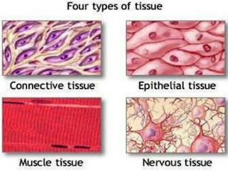
Types of Tissue

1.     Epithelium Tissue:

·       (EpI-Upon Thelium-membrane)

·       This tissue grows upon a basement membrane.

·  Epithelial layers contain no blood vessels, so they must receive nourishment via diffusion of substances from the underlying connective tissue, through the basement membrane.

Classification of Epithelium Tissue:

A.    Based on number of layers:

·       Simple Epithelium: It is a single layered or one cell thick. Every cell in direct contact with the underlying basement membrane

·       Stratified Epithelium: It is multi-layered.

B.    Morphology of their cells:

·       Squamous epithelium (flat and scale like cells)

·       Cuboidal epithelium (Cube-shaped cells)

·       Columnar epithelium (Column-Shaped cells)

Epithelium tissue is present in outer layer of skin, inner lining of mouth, digestive tract, visceral organ, blood vessels and glands.

Functions of Epithelium Tissue

·       This tissue is responsible for absorption of water & nutrient, elimination of waste, secretion of enzymes, hormones and protection.


2.     Connective Tissue: Connective tissue that connects supports, bind or separates other tissues or organs. All connective tissue consists of three main components:

·       The cells of connective tissue include fibroblast, adipocytes, macrophages, mast cells etc.

·       The fibers: elastic, collagenous fibers.

·       The ground substances (matrix)

Types of Connective tissue:

There are three types of connective tissue:

a)     Fibrous connective tissue: e.g. Adipose tissue, Areolar tissue

·       Ligaments: the fibrous connective tissue which joint bone to bone strongest ligaments of human body is illio femoral ligament which joint hip joint (ilium) to femur in front of the joint.

·       Tendon: The fibrous connective tissue which joint bone to muscle. Strongest tendon of human body is Achilles tendon of heal cord or calcaneal tendon, situated at back of the leg, which joint the heal bone to calf muscle.

b)    Skeletal connective tissue: It include bone and cartilage

·       Osteoblast: bone forming cell.

·       Osteoclast: bone destructing or shaping cell.

·       Chondroblast: cartilage forming cell.

v The fibrous connective tissue and skeletal connective tissue provide the mechanical support to human body.

c)     Fluid connective tissue:

·       It includes blood, plasma and lymph.

·       The matrix of fluid connective tissue is in liquid state.


3.     Muscular Tissue: muscular tissue is soft tissue that composes muscle and gives rise to muscle ability to contract.

·       Myocyte: Muscle forming cell.

·       Myogenesis: The process of muscle formation.

Types of Muscular tissue:

There are three types of muscle in human body:

a)     Skeletal muscle: they are striated in structure and under voluntary control is situated in association with skeletal muscle are multinucleated usually. The skeletal muscles form 40 to 50% of body mass.

·       Sarcomere: it is defining as the structural and functional unit of skeletal muscle. It is also called the basic contractile unit of muscle.

·       Functions: they carry out movement of the body, maintain the posture of the body and support the body.

b)    Smooth muscle: Smooth muscle is situated in association with visceral, it is also called visceral muscle. They are Non-striated and Involuntary in nature.

·       Function: they are responsible for the contractility of hollow organs, such as blood vessels, GI Tract, bladder etc.

c)     Cardiac Muscle: They forms the musculature (myocardium) of the heart. These muscles are striated and Involuntary in nature.

·       Function: cardiac muscles are the self –contacting, automatically regulated and must continue to contract in rhythmic fashion for the whole life of the human.

4.     Nervous Tissue: Nervous tissue consists of Neurons and supporting cells.

·       Neuron or nerve cell is defined as the structural and functional unit of nervous system.

·       Neuron is similar to any other cell in the body having nucleus and all the organelles in cytoplasm, however, it is different from other cell by two ways:

                      i.          Neuron has branches or processes called Axon and Dendrites.

                    ii.          Neuron does not have centrosome, so it cannot undergo division that’s why have minimum power or regeneration.

Types of Nervous tissue:

On the basis of function, nerve cells are classified in to two types:

a)     Sensory or afferent Neurons: They carry the sensory impulses from periphery to CNS.

b)    Motor or Efferent neurons: They carry the motor impulses from CNS to periphery.

v Synapse: The junction by which the neurons communicate with each other.

Anatomy and Physiology Quiz Series 

Coronary Artery Disease 


SHOCK

 A clinical syndrome marked by inadequate perfusion and oxygenation of cells, tissues and organs, usually as a result of severe or markedly lowered blood pressure. The shock syndrome is a life-threatening medical emergency and requires very careful therapy and monitoring.

Type of Shock: Based on causes of shock

1. Cardiogenic shock-

  • Failure of the heart to pump an adequate supply of blood and oxygen to body tissues.
  • The most common cause of cardiogenic shock is acute myocardial infarction.
2. Anaphylactic shock-

  • Rapidly developing, systemic anaphylaxis that produces life-threatening acute airway obstruction and vascular collapse within minutes after exposure to an antigen.
  • The most common cause is a type I allergic or hypersensitivity reaction.
  • The most common agents are bee or wasp venoms, drugs (e.g. penicillin) and radiographic contrast media.
3. Hypovolemic shock-

  • Shock occurring when there is an insufficient amount of fluid in the circulatory system.
  • Most common causes are excessive bleeding, diarrhea, vomiting, fluid loss from fistulas or burns and third space shifting of fluid into the abdominal cavity (ascites) or other intestinal tissue (edema).
4. Neurogenic shock-

  • A form of shock due to decreased peripheral vascular resistance caused by damage to either the brain or the spinal cord.
  • The decreased peripheral vascular resistance (vasodilation) and subsequent pooling of blood within peripheral vessels results in hypotension and warm, dry or pink skin.
5. Septic shock-

  • Shock as the result of sepsis (the presence of pathogens in the bloodstream).
  • The most common organism are gram-negative (common) and gram-positive bacteria, but fungi and other organisms may also be responsible.
6. Hypoglycemic shock or insulin shock-

  • Shock produced by extremely low blood sugars (e.g. less than 40mg/dl) usually caused by an injection of an excessive amount of insulin, failure  to eat after an insulin injection and insulinomas (insulin secreting tumors which cause hypoglycemia).
  • Insulin-related hypoglycemic shock may be intentionally induced in the treatment of certain psychiatric conditions.
Clinical Manifestation:

  • Shock results in failure of multiple organ systems including the brain, heart, kidney , lungs, skin and gastrointestinal tract.
  • Subjective (symptom): Restlessness, Paresis of extremities, Confusion, Syncope
  • Objective (sign): Weak, rapid, thready pulse (tachycardia); Shallow, rapid respiration (tachypnea); Diaphoresis; Cold, clammy and cyanotic skin (except neurogenic shock); Pallor; Decreased  urine output (oliguria); Progressive loss of consciousness; Decreased mean arterial pressure (normal is 80 to 120 mm Hg).
Management:

  • The shock syndrome  is a life-threatening medical emergency and requires very careful therapy and monitoring 
  • Attempts to restore normal blood pressure and tissue perfusion include:-
            - Fluid resuscitation (in hypovolemic shock)

            - Control of hemorrhage (in shock caused by trauma or bleeding).

            - Administer corticosteroids and antihistamine such as chlorpheniramine maleate,                  cetirizine etc. in anaphylactic shock.

            - Administration digoxin for pressor support (in cardiogenic) and antibiotic (in septic                shock).

            - Administration of epinephrine (in anaphylaxis or neurogenic shock for peripheral                     vasoconstriction).

            - Administration oral or parenterally sugars, typically glucose (in hypoglycemia or                 insulinomas).  

  • Insert indwelling urinary catheter to track urine output hourly.
  • SaO2, arterial blood gas levels (ABGs), and ventilatory functions are monitored to determine the need for ventilatory support.
  • Suitable position used during shock is trendelenburg position.


BLOOD CLOTTING FACTORS

The blood clotting factors in human body are as follows:

  1. Factor I       : Fibrinogen 
  2. Factor II      : Prothrombin (deficiency cause thrombophilia)
  3. Factor III    : Tissue factor (Thromboplastin)
  4. Factor IV    : Calcium 
  5. Factor V      : Labile factor (proaccelerin)
  6. Factor VI    : Unassigned/presence does not proved
  7. Factor VII   : Stable factor or proconvertin
  8. Factor VIII : Antihemophilic factor A (deficiency cause hemophilia A)
  9. Factor IX    : Antihemophilic factor B or Christmas factor (deficiency cause hemophilia B)
  10. Factor X     : Stuart - Power factor
  11. Factor XI   : Hegeman factor
  12. Factor XII : Fibrin stabilizing factor 


Head Injury

Cerebrovascular Accident

Brain Anatomy

Appendectomy

Definition
  • The excision of the appendix usually performed to remove an acutely inflamed organ.
  • Many surgeons perform an appendectomy as a prophylactic procedure when operating in the abdomen for other reasons. This procedure is then referred to as an incidental appendectomy.

Position
  • Supine, with arms extended on armboards

Incision Site
  • McBurney (muscle splitting) incision.

Packs/ Drapes
  • Laparotomy pack
  • Four folded towels

Instrumentation
  • Major Lap tray or minor tray
  • Internal stapling device

Supplies/ Equipment
  • Basin set
  • Blades
  • Needle counter
  • Penrose drain
  • Culture tubes
  • Solutions
  • Sutures
  • Internal stapling instruments
  • Medication

Procedure
  1. An incision is made in the right lower abdomen, either transversely oblique (McBurney) or vertically (for a primary appendectomy).
  2. The surgeon’s assistant retracts the wound edges with a Richardson or similar retractor.
  3. The appendix is identifies and its vascular supply ligated.
  4. The surgeon grasps the appendix with a Babcock clamp, and delivers it into the wound site.
  5. The tip of the appendix may then be grasped with a Kelly clamp to hold it up, and a moist Lap sponge is placed around the base of the appendix (stump) to prevent contamination of bowel contents, in case any spill out occurs during the procedure.
  6. The surgeon isolates the appendix from its attachments to the bowel (mesoappendix) using a Metzenbaum scissors.
  7. Taking small bits of tissue along the appendix, the mesoappendix is double-clamped, and ligated with free ties.
  8. The base of the appendix is grasped with a straight Kelly clamp, and the appendix is removed.
  9. The stump may be inverted into the cecum, using a purse-string suture on a fine needle, cauterize with chemicals, or simply left alone after ligation.
  10. Another technique is to devascularize the appendix and invert the entire appendix into the cecum.
  11. The appendix, knife, needle holder, and any clamps or scissors that have come in contact with the appendix are delivered in a basin in the circulating nurse.
  12. The wound is irrigated with warm saline, and is closed in layers, except when an abscess has occurred, as with acute appendicitis.
  13. A drain may be placed into the abscess cavity, exiting through the incision or a stab wound.
  14. An alternative technique may be use the internal stapling device, by placing the stapling instrument around the tissue at the appendiocecum junction.
  15. By using the technique, the possibility of contamination from spillage is greatly reduced.

Perioperative Nursing Consideration
  1. Instruments used for amputation of the appendix are to be isolated in a basin.
  2. If ruptured, the case must be considered contaminated, and the surgeon may elect to use antibiotic irrigation prior to closure of the abdomen with an insertion of a drain.
  3. There may be no skin closure of the wound if the appendix has rupture.

Anatomical Body Landmark

Anatomical Position

The first important thing in learning about anatomy and physiology is to know the anatomical position and body landmarks. To accurately describe body parts and position, initial reference point and directional terms should be used in a medical setting. The correct anatomical position is standing up (erect) with the feet parallel and the arms hanging at the sides with the palms facing forward and the thumbs pointing away from the body.

Body Landmarks

To precisely point out the chief complaint of a patient, the nurse or physician uses anatomical terms representing a certain body part. For example, a patient walks in the emergency room with a hacking wound on the posterior portion of the left lower leg. To clearly state the area of injury the nurse uses the term “sural” which means the posterior surface of the lower leg rather than writing “back area of the lower leg”. Knowing these terms not only give the nurse a more accurate formulation of chief complaints but it also saves time of a good deal of description.


Anterior Body Landmarks

Abdominal – anterior body trunk inferior to ribs
Acromial – point of shoulder
Antecubital – anterior surface of elbow
Axillary – armpit
Brachial – arm
Buccal – cheek area
Carpal – wrist
Cervical – neck region
Coxal – hip
Crural – leg
Digital – fingers, toes
Femoral – thigh
Fibular – lateral part of the leg
Inguinal – area where thigh meets body part
Nasal – nose area
Oral – mouth
Orbital – eye area
Patellar – anterior knee
Pelvic – area overlying the pelvis anteriorly
Pubic – genital region
Sternal – breastbone area
Tarsal – ankle region
Thoracic – chest
Umbilical – navel

Posterior Body Landmark
Calcaneal – heel of foot
Cephalic – head
Deltoid – curve of shoulder formed by large deltoid muscle
Femoral – thigh
Gluteal – buttock
Lumbar – area of back between ribs and hips
Occipital – posterior surface of head
Olecranal – posterior surface of elbow
Popliteal – sacral
Scapular – shoulder blade region
Sural – posterior surface of the lower leg
Vertebral – area of spine
Plantar – sole of the foot
Directional Terms
To clearly explain exactly the relation of a body structure to each other, directional terms are used. For example to describe the relationship or location of the heart to the arms, we can say “the heart is located in between the arms”. Using anatomical terminology, this is expressed as “the heart is medial to the arms.” Hence, it is a more clear and precise statement.
Superior – above
Inferior – below
Anterior – in front of
Posterior – behind
Medial – middle
Lateral – away from the middle; at outer the sides
Intermediate – between a more medial and a more a lateral surface
Proximal – close to the body part
Distal – away from a body part
Superficial – external; at the surface
Deep – internal; away from the surface

ABNORMALITIES OF MOTOR BEHAVIOUR

  • Psychomotor Retardation: Slowed mental and motor activities.
  • Stupor: A state in which a person does not react to the surroundings: (mute, immobile and unresponsive).
  • Catatonic Stupor: Stupor with rigid posturing
  • Psychomotor Agitation: Restlessness with psychological tension. (Patient is not fully aware of restlessness.)
  • Catatonic Excitement: Marked agitation, impulsivity and aggression without external provocation.
  • Chorea: sudden involuntary movement of several muscle groups with the resultant action appearing like part of voluntary movement.
  • Aggression: Verbal or physical hostile behaviour, with rage and anger.
  • Akathisia: Inability to keep sitting still, due to a compelling subjective feeling of restlessness.
  • Dyskinesia: Restless movement of group of muscles (face, neck, hands).
  • Dystonia: Painful severe muscle spasm.
  • Torticollis: Contraction of neck muscles.
  • Tics: Sudden repeated involuntary muscle twisting. e.g. repeated blinking, grimacing.
  • Compulsions: Compelling repeated irrational actions associated with obsessions. e.g. repeated hand washing.
  • Echopraxia: Imitative repetition of movement of somebody.
  • Waxy Flexibility: Patient’s limbs may be moved like wax, holding position for long period of time before returning to previous position.
  • Stereotypies: Purposeless repetitive involuntary movements. e.g. foot tapping, thigh rocking.
  • Mannerism: Odd goal-directed movements. e.g. repeated hand movement resembling a military salute.
  • Automatic obedience:  the pt. carries out every instruction regardless of the consequences.
  • Perseveration: is a senseless repetition of a goal-directed action, a particular response, such as a word, phrase, or gesture which has already served its purpose (beyond their relevance).
  • Dyspraxia; inability to carryout complex motor tasks, although the component motor movements are preserved.
  • Omega sign (Athanassio): the occurrence of a fold like the Greek letter omega in the forehead above the root of the nose produced by the excessive action of the corrugator muscle; seen in depression.
  • Ambitendency: a motor symptom of schizophrenia in which there is an alternating mixture of automatic obedience and negativism.
  • Mitgehen: The pt. moves his body in the direction of the slightest pressure on the part of the examiner. seen in catatonia
  • Mitmachen (Co-operation): The body can be put to any position without any resistance on the part of the pt. seen in catatonia.
  • Trichotillomania: a condition characterized by an overwhelming urge to pluck out specific hairs.
  • Pyromania: impulsive repetitive, deliberate fir-setting without external rewards.
  • Dipsomania: uncontrollable craving for alcohol or compulsive drinking of alcohol.
  • Kleptomania: a disorder in which the individual impulsively steals things other than personal use or financial gain.
  • Negativism:  an apparently motiveless resistance to all commands and attempts to be moved or doing just the opposite.
References
  1. Psychiatry, Third Edition. Edited by Allan Tasman, Jerald Kay, Jeffrey A. Lieberman, Michael B. First and Mario Maj. John Wiley & Sons, Ltd, 2008.
  2. Sims, A. Symptoms in the Mind: An Introduction to Descriptive Psychopathology (3rd ed). Elsevier, 2002.
  3. Fish, F. Clinical Psychopathology, Signs and Symptoms in Psychiatry. Bristol: J. Wright & Sons. 1967.